keskiviikko, 19. elokuu 2026

In vivo CAR-T vs Traditional CAR-T: Reshaping the Future of Cell Immunotherapy

Overview


On November 2, commercial insurance catalog negotiations officially began, with five CAR-T therapies entering the negotiation list: Fosun Kite’s Axicabtagene Ciloleucel, JW Therapeutics’ Relmacabtagene Autoleucel, IASO Bio’s Equecabtagene Autoleucel, CARsgen Therapeutics’ Zevo-Cabtagene Autoleucel, and Abogen’s Nacacabtagene Autoleucel.


As a supplement to the basic medical insurance system, the innovative drug catalog under commercial insurance adopts a negotiated pricing mechanism instead of deep price cuts, reserving space for the development of innovative therapies.


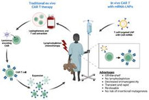
Since the beginning of this year, the biopharmaceutical industry has witnessed an intense “arms race.” AstraZeneca, AbbVie, Gilead, and BMS have each spent hundreds of millions of dollars to acquire early-stage in vivo CAR-T companies. In sharp contrast, traditional autologous and allogeneic (off-the-shelf) CAR-T therapies are showing growth bottlenecks — complex manufacturing, high cost, and limited efficacy against solid tumors.
 

a02458fcb8f8b0ef00b808d32bce83e8.png

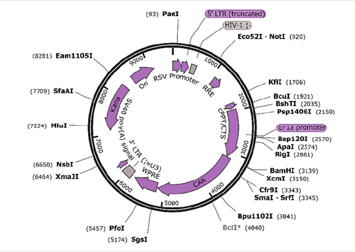
Schematic diagram of CAR lentiviral vector structure


Reconstructing the Treatment Logic: Precision Delivery and Cellular Engineering


Although traditional ex vivo CAR-T therapy shows great potential, it has always been limited by three major challenges:


Complex process: 


It requires blood collection, ex vivo T-cell modification and expansion, which are time-consuming and rely on specialized facilities.


Limited manufacturing capacity: 


The personalized nature of production makes large-scale manufacturing difficult, restricting patient accessibility.


Preconditioning dependency: 


Patients need to undergo chemotherapy to deplete immune cells before infusion, increasing infection risks.


In contrast, in vivo CAR-T delivers CAR genes directly into the body via specific carriers, precisely targeting T cells, NK cells, or even hematopoietic stem cells for in situ genetic modification. This strategy retains the precision killing power of CAR molecules while achieving scalability — breaking through the long-standing limitations of traditional CAR-T therapies.

Technical Routes:

“Long-acting” Lentiviral Vector VS “Controllable” LNP Vector

Lentiviral vectors – One-time treatment for long-term protection:


Lentiviral vectors integrate the CAR gene stably into the T-cell genome, enabling durable CAR expression and prolonged tumor-killing capability. The core technologies lie in T-cell–specific targeting, transduction efficiency, and safety regulation.
 

01. T-cell targeting transformation: from broad tropism to T-cell specificity


Traditional lentiviruses use broadly tropic envelope proteins (such as VSV-G), which can infect hepatocytes and endothelial cells. Dual engineering — detargeting + retargeting — achieves T-cell–specific delivery:

Envelope protein engineering: 

Replace VSV-G with a CD28L chimeric envelope, fusing the T-cell co-stimulatory ligand CD28L. This specifically binds to the CD28 receptor on T cells, increasing transduction efficiency 30-fold over wild-type lentivirus while reducing off-target hepatocyte infection to <0.1%.

Genomic element optimization: 

Introduce an RACR/CAR system where the lentiviral vector carries both a “reverse responsive element” and the CAR gene. Without drug administration, CAR expression remains silent and T cells are quiescent. After oral administration of the small molecule AP21967, the responsive element changes conformation to activate CAR expression, peaking in 48 hours and silencing again within 7 days after drug withdrawal — reducing CRS incidence by 60%.

02. Transduction efficiency optimization: high-efficiency T-cell modification at low doses


Lentiviral transduction efficiency depends on viral titer, T-cell activation, and co-stimulatory signals. The following strategies enable breakthroughs:

High-titer production: 

By adopting a “suspension + serum-free culture” system and optimizing HEK293T transient transfection, viral titers increased from 1×10⁸ TU/mL to 5×10⁹ TU/mL. This supports effective transduction at one-tenth the standard dose, minimizing off-target risks.


T-cell activation synergy: 

Embedding an IL-2 mimetic peptide within the vector activates T-cell proliferation signals, increasing the proportion of G1-phase T cells from 35% to 62%, boosting transduction efficiency 1.8×. Meanwhile, integrating 4-1BBL into the envelope provides a second co-stimulatory signal by binding 4-1BB on T cells, enhancing survival and persistence — with CAR-T cells persisting up to 12 months in cynomolgus monkey models.

03. Clinical safety regulation: enabling long-lasting T-cell killing

Hematologic malignancies — Sustained response at low dose: 

With CD45-ligand lentiviral delivery of CD19 CAR at 1×10⁸ TU/kg, peripheral T-cell transduction reached 63%, tumor burden dropped by 92% in lymphoma mouse models, and IL-6 levels were 50% lower than ex vivo CAR-T.

Solid tumors — Enhanced tumor penetration: 

Lentiviral vectors co-delivering “BCMA-CAR + TGFβ-neutralizing peptide” in multiple myeloma models neutralized TGFβ-mediated immunosuppression, enabling CAR-T infiltration deep into tumor cores. The Treg proportion in tumor microenvironment decreased from 32% to 11%, improving killing efficiency by 2.5×.

LNP Vector – “Safe, Controllable, and Flexible”


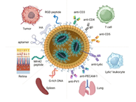
CAR mRNA does not integrate into the genome, naturally avoiding genotoxic risks. mRNA-LNP uses “antibody-targeted LNPs” to deliver CAR mRNA directly to T cells in vivo, achieving in situ T-cell reprogramming. The core technologies involve Ab-LNP targeting, formulation, and clinical validation.

01. Ab-LNP specific targeting: precise recognition of T cells

The key to Ab-LNP lies in antibody modification that forms a specific bridge between LNPs and T-cell receptors. The process includes: antibody conjugation of CD3/CD8-specific antibodies to LNP surfaces → specific binding to T-cell receptors → endocytosis and mRNA release → high-efficiency CAR expression within cytoplasm.
 

IMG_256

02. Ab-LNP formulation: enhancing T-cell transduction efficiency

Ab-LNP preparation optimizes antibody density, ionizable lipid pKa, particle size, and mRNA loading to maximize transduction efficiency.

03. Ab-LNP clinical validation: safety and efficacy

mRNA-LNP–based in vivo CAR-T technology has achieved multiple breakthroughs:

  • Over 90% clearance of hematologic malignancies without hepatic toxicity;
  • Significant symptom relief in autoimmune diseases (e.g., lupus) with no neurotoxicity;
  • Effective solid tumor control by incorporating chemokine mRNA to improve infiltration — combined with PD-1 inhibitors, tumor suppression reached 89%.


Risks and Outlook: The “Last Mile” from Lab to Bedside


Although in vivo CAR-T holds great promise, several barriers remain before clinical translation:

Lentiviral long-term risks: 

Persistent CAR expression may cause chronic B-cell depletion; genomic integration risks require long-term monitoring.

mRNA-LNP short-lived expression: 

Transient CAR expression limits long-term tumor control, requiring repeat dosing.

Cell selectivity: 

Off-target transduction of non-intended cells can reduce efficacy or cause organ toxicity.

The industry is working to overcome these issues: lentiviral vectors are being optimized with improved envelope design to reduce immunogenicity and integration risks, while mRNA-LNP systems are leveraging circular mRNA to extend expression duration. Both are evolving toward more precise targeting and longer-lasting therapeutic effects.

In vivo CAR-T therapy marks a key evolutionary step in cell therapy — shifting from personalized, case-by-case treatment to standardized and scalable therapeutic models.
This technological revolution not only addresses the cost and accessibility challenges of traditional CAR-T therapy but also heralds a new era in cell-based immunotherapy.

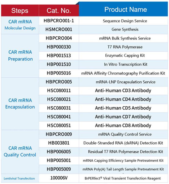
Related Products
 

3221a398be35f8eadc5b705cd55a6ed4.png

Bibliography


1.Du L, Nai Y, Shen M, Li T, Huang J, Han X, Wang W, Pang D, Jin A. IL-21 Optimizes the CAR-T Cell Preparation Through Improving Lentivirus Mediated Transfection Efficiency of T Cells and Enhancing CAR-T Cell Cytotoxic Activities. Front Mol Biosci. 2021 Jun 4;8:675179.

2.Dai Q, Han P, Qi X, Li F, Li M, Fan L, Zhang H, Zhang X, Yang X. 4-1BB Signaling Boosts the Anti-Tumor Activity of CD28-Incorporated 2nd Generation Chimeric Antigen Receptor-Modified T Cells. Front Immunol. 2020 Nov 13; 11: 539654. 

3.Xu J, Liu L, Parone P, Xie W, Sun C, Chen Z, Zhang J, Li C, Hu Y, Mei H. In-vivo B-cell maturation antigen CAR T-cell therapy for relapsed or refractory multiple myeloma. Lancet. 2025 Jul 19;406(10500):228-231. 

4.Andorko JI, Russell RM, Schnepp BC, Grubaugh D, Mullen KF, Wakabayashi A, Carrington LJ, O'Malley T, Kuri-Cervantes L, Culp TD, Johnson PR. Targeted in vivo delivery of genetic medicines utilizing an engineered lentiviral vector platform result in CAR T and NK cell generation. Mol Ther. 2025 Oct 1;33(10):4937-4952. 

5.Jacob Garcia, Christine Dehner, Jeffrey Teoh, Wayne Wallis, A Phase 1, Multicenter, Open-Label Study of UB-VV111 in Combination with Rapamycin in Relapsed/Refractory CD19+ B-Cell Malignancies, Blood, Volume 144, Supplement 1, 2024, Page 1750. 1, ISSN 0006-4971.

keskiviikko, 19. elokuu 2026

HZYMES Enzyme Preservation and Lyophilization Technology: Ensuring Stability and Efficiency in Biote

Enzymes play a pivotal role in countless industrial applications, from pharmaceuticals to food processing and environmental management. However, one of the primary challenges in working with enzymes is their stability. Enzymes are delicate proteins, and their functionality can degrade over time due to environmental factors such as temperature fluctuations, changes in pH, and the presence of contaminants. Therefore, effective preservation methods are essential to ensure their long-term activity and usability.


Among various preservation techniques, enzyme lyophilization (freeze-drying) stands out as a highly effective solution, offering enhanced stability and ease of storage. This article explores the science behind enzyme preservation and the key role that lyophilization technology plays in ensuring enzyme stability for industrial applications.


II. Enzyme Preservation: Why It Matters


Enzyme stability is critical for industries that rely on their catalytic properties, such as pharmaceuticals, food production, and bioenergy. If enzymes lose their activity during storage or transportation, the entire production process can be affected, leading to operational delays and financial losses. Additionally, the cost of replacing enzymes or maintaining a cold supply chain for liquid enzymes can be prohibitive.


The need for efficient enzyme preservation methods is therefore clear. Traditional methods, such as storing enzymes in liquid form under refrigeration, have limitations in terms of shelf life, ease of transportation, and cost. Lyophilization, which involves removing water from the enzyme solution to form a stable dry product, addresses these issues while preserving enzyme activity for long periods.


III. What is Lyophilization (Freeze-Drying)?


Lyophilization is a specialized process used to preserve sensitive biological materials, such as enzymes, by removing water through sublimation. In this process, an enzyme solution is first frozen at a low temperature, and then the ice is removed via a vacuum in a primary drying phase (sublimation), followed by secondary drying to remove any residual moisture.


The main advantage of lyophilization is that it allows enzymes to be stored in a dry, stable form that is easy to handle and transport. By preserving the enzyme in a solid form, lyophilization significantly extends its shelf life while maintaining its biological activity. This makes lyophilized enzymes ideal for long-term storage and global distribution, especially in industries where cold storage is not feasible.


IV. The Advantages of Enzyme Lyophilization


The lyophilization of enzymes offers a variety of advantages:


 Enhanced Stability: 


Lyophilized enzymes are far more stable than their liquid counterparts. By removing water, the enzyme’s molecular structure is protected, reducing the risk of denaturation or degradation that occurs in the presence of moisture or fluctuating temperatures.


• Longer Shelf Life: 


Lyophilized enzymes can be stored for extended periods (often years) without significant loss of activity, making them ideal for industries that require long-term enzyme storage or that need to keep inventory on hand for emergencies.


 Ease of Transport: 


Lyophilized enzymes do not require refrigeration, making them easier and less costly to transport. This is particularly important for global supply chains, where temperature-controlled logistics can be both expensive and complicated.


 Cost-Effectiveness: 


While the initial cost of lyophilization equipment may be high, the long-term savings in storage and transportation costs make lyophilized enzymes an economically viable solution for many industries.


V. Key Factors Affecting Enzyme Lyophilization


The lyophilization process must be carefully controlled to ensure enzyme stability. Several factors affect the success of enzyme freeze-drying:
 

 Enzyme Formulation: 


The addition of stabilizing agents, such as cryoprotectants or buffering agents, is critical in preserving enzyme activity during the freeze-drying process. These substances help to protect the enzyme’s structure from damage caused by freezing or dehydration.


 Lyophilization Conditions: 


The temperature, vacuum pressure, and drying time must be optimized to prevent denaturation. The freezing phase must ensure that the enzyme’s structure is preserved, while the drying phases need to be gentle to avoid destabilizing the enzyme’s active sites.


 Enzyme Structure: 


Enzymes with more complex structures or those requiring specific conditions for activity may be more challenging to lyophilize successfully. The choice of enzyme formulation and stabilizers plays a crucial role in overcoming these challenges.


VI. Lyophilization in Different Enzyme Applications


Enzyme lyophilization has proven to be beneficial in numerous industrial sectors:


 Pharmaceutical Industry: 


In pharmaceutical applications, lyophilized enzymes are used in drug formulations, diagnostics, and enzyme replacement therapies. The stability and portability of lyophilized enzymes ensure that they maintain their efficacy throughout the supply chain.


 Food Industry: 


Enzyme preparations for food processing, such as those used in brewing, dairy, or baking, are often lyophilized to maintain high activity levels without refrigeration. Lyophilization ensures that enzymes can be stored for extended periods, allowing for more efficient production processes.


 Bioenergy: 


Enzyme lyophilization also plays a role in biofuels, where enzymes are required for biomass breakdown and fermentation. The ability to store and transport these enzymes in a stable, dried form ensures a continuous and efficient biofuel production process.


 Health and Beauty: 


Lyophilized enzymes are used in the health and beauty industry for various purposes, including skincare formulations. The ability to preserve enzymes in a potent form makes them valuable ingredients in a variety of cosmetic products.


VII. Advancements in Lyophilization Technology


The technology behind enzyme lyophilization has advanced significantly over the years. Some of the key innovations include:


 Advanced Freeze-Drying Equipment: 


Modern lyophilization equipment offers precise control over temperature, pressure, and drying times, resulting in better-preserved enzymes with higher activity retention.


 Enhanced Stabilizers: 


New stabilizing agents, including synthetic and natural protectants, have been developed to improve the success of lyophilization, especially for more fragile enzymes.


 Nanotechnology: 


Research into nanoencapsulation and other advanced materials has opened the door to even more efficient enzyme preservation, allowing for the encapsulation of enzymes in nanoparticles that protect them during lyophilization and enhance their stability after rehydration.


VIII. Challenges and Limitations of Enzyme Lyophilization

Despite its many advantages, enzyme lyophilization does have some challenges:

 Enzyme Denaturation: 

Even with optimal conditions, some enzymes may lose activity during the freeze-drying process. Identifying the best formulation and stabilizers is crucial to minimizing this risk.

 Rehydration Time: 


Some lyophilized enzymes may take longer to rehydrate than others, which can be a limitation for certain high-volume industrial processes.

 Cost of Technology: 


The initial cost of lyophilization technology and the need for ongoing maintenance can be prohibitive for smaller businesses, although the long-term benefits often outweigh the initial investment.


IX. Case Study: Successful Lyophilization of Enzymes


One real-world example of successful enzyme lyophilization can be seen in the pharmaceutical industry. HZYMES, a leading biotech company, developed a lyophilized enzyme solution for an enzyme replacement therapy used in the treatment of a rare metabolic disorder. By employing advanced lyophilization techniques, HZYMES was able to extend the shelf life of the enzyme by over 50% compared to previous methods. This not only reduced transportation costs but also improved patient access to the treatment across global markets.

Conclusion

Enzyme preservation and lyophilization technology are critical components of the biotechnology and industrial sectors. Lyophilization offers significant advantages in terms of enzyme stability, ease of transport, and cost-effectiveness. As the demand for high-quality enzymes continues to grow across diverse industries, advancements in freeze-drying technology will further enhance enzyme preservation, allowing for more efficient and sustainable production processes.


For companies like HZYMES, embracing and advancing enzyme lyophilization technology ensures that enzymes remain potent, accessible, and ready for use, driving innovation and success in a variety of industries.

keskiviikko, 19. elokuu 2026

One Plasmid, One Week, One Revolution: How T7-ORACLE Rewrites Protein Evolution

Background


No complex instruments. No repetitive manipulation. Just one plasmid—and your target gene can undergo continuous hypermutation inside living cells, enhancing protein activity by thousands of times within a week!


It may sound like a synthetic biologist’s dream, but it has now become reality. On August 7, 2025, Peter Schultz’s team at the Scripps Research Institute published a groundbreaking study in Science, unveiling a system called T7-ORACLE, an orthogonal replication system that enables continuous, rapid, and targeted gene hypermutation in E. coli. This represents a revolutionary breakthrough in protein-directed evolution.


34f71ac3e9776ef5e7d117efe2b40ad1.png


Bottlenecks in Traditional Evolution Technologies


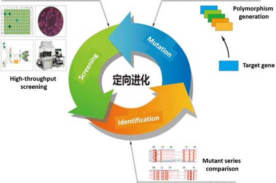
Directed evolution is a key strategy for optimizing protein function, yet traditional approaches such as error-prone PCR or DNA shuffling have significant limitations: they rely on multiple rounds of in vitro mutation–transformation–screening cycles, which are labor-intensive and time-consuming, and they often fail to achieve deep evolutionary changes.


Although recent in vivo evolution technologies (like PACE) have improved efficiency, they still face issues such as non-targeted mutations, host genome instability, and the need for specialized equipment.


Inspired by bacteriophage T7, the Schultz team developed a fully orthogonal replication system composed of multiple components: T7 replicative proteins, including RNA polymerase, DNA polymerase, helicase–primase fusion protein, and single-stranded DNA-binding protein.


Mechanism:


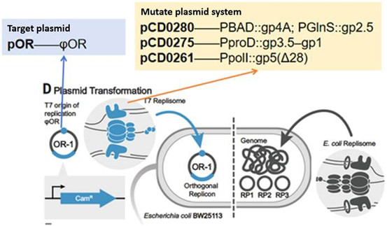
The system works by expressing T7 RNA polymerase, DNA polymerase, helicase–primase, and single-stranded binding protein, which specifically recognize and replicate circular target plasmids carrying the T7 replication origin.


To promote replication, a catalytically defective T7 lysozyme is fused to the T7 RNA polymerase. The inhibitory effect of T7 lysozyme prevents full transcription, generating short RNA primers that initiate replication by T7 DNA polymerase.


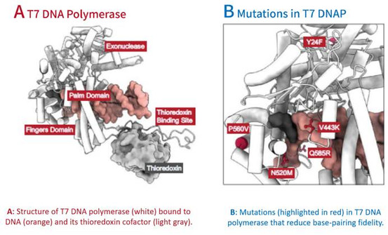
By introducing engineered T7 DNA polymerase variants (Δ28 deletion and key mutations N520M, P560V, and V443K), the fidelity of replication is drastically reduced.


a252ad172be326d678e26a999d4073bd.png

Figure 1. Schematic diagram of the directed evolution workflow


T7-ORACLE: A Precise “Genetic Editing Engine”


The Schultz team designed the T7-ORACLE system as a fully orthogonal replication mechanism inspired by bacteriophage T7.


It consists of several key components: T7 RNA polymerase, DNA polymerase, helicase–primase fusion protein, and single-stranded binding protein.


The system specifically targets plasmids carrying the T7 replication origin (φOR) for replication.


3429b2f94ab08b89478c3a18a412d87d.png

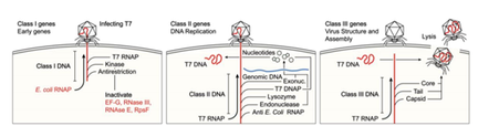
Figure 2. Life cycle of T7 bacteriophage


Class I genes encode host-suppression functions and T7 RNA polymerase;
Class II genes encode the T7 replication machinery;
Class III genes encode T7 capsid and lysis proteins.


1818bdd7d670901ff382543cc9705fb1.png

Figure 3. Schematic diagram of system design principle


To maximize mutational efficiency, the engineered DNA polymerase exhibits reduced proofreading activity and base selectivity, resulting in controlled hypermutation.


Most importantly, this system targets only the plasmid-borne genes without disturbing the host genome, allowing E. coli cells to maintain normal growth while achieving extremely high mutation rates in the target sequence.


How Is a Million-Fold Increase in Mutation Rate Achieved?


The researchers used rational design and directed evolution to iteratively optimize T7 DNA polymerase:


Removing proofreading activity: 


An exonuclease-deficient mutant (Δ28) raised the mutation rate 25-fold.


Reducing base-pairing fidelity: 


Key mutations N520M, P560V, and V443K further lowered replication accuracy.


279b519ed00c948bb375c62c5fdb1e39.png

Figure 4. Engineered T7 DNA polymerases involved in this study


The resulting five-mutation polymerase achieved a mutation rate of 1.7×10⁻⁵ per base per generation, nearly 100,000 times higher than the host genome mutation rate, while maintaining high transformation efficiency (2.4×10¹⁰ cfu/μg).


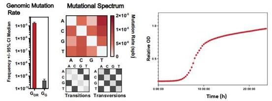
Stability tests of the optimized system included:


Fluctuation analysis


 confirming excellent orthogonality (genomic mutation rate 4.4×10⁻¹⁰ spb);


Sequencing of mutation spectra


showing unbiased substitution patterns (balanced transition/transversion rates);


Serial passage experiments


confirming plasmid stability and negligible metabolic burden for plasmids up to 13 kb


28bf5f60993a8dc17527842d72fe0c32.png

Figure 5. Stability testing of the optimized system


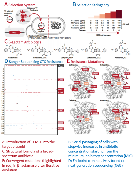
Application: Hyperactive β-Lactamase Evolved in Just One Week


To validate the system’s practicality, the team applied it to evolve TEM-1 β-lactamase.


In less than one week, mutants exhibiting 5,000-fold higher resistance to several clinically important antibiotics (aztreonam, cefotaxime, ceftazidime, and cefepime) were obtained.


Remarkably, the mutations that emerged (such as G238S, R164H, and E104K) were identical to those observed in clinical antibiotic-resistant strains, proving that the system accurately mimics natural evolutionary processes.


9c2d4d897a985b1e39ca9e5fd36919d2.png

Figure 6. Continuous evolution of β-lactamase


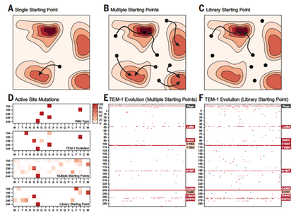
Breaking Local Optima: From a Single Starting Point to Global Library Evolution


One of T7-ORACLE’s major advantages is its compatibility with pre-constructed mutation libraries.


The researchers compared different evolutionary starting points:


From the wild-type sequence 


– the evolution pathway was highly convergent (single-directional evolution).


From single-point saturation mutants 


– multiple unidirectional trajectories emerged.


From a full saturation library 


– multiple multidirectional trajectories were observed, enabling exploration of the global fitness landscape and identification of global optima.


403956b15d2b1b971faaf6b9e1d90264.png

Figure 7. Exploring broader fitness landscapes from multiple starting points toward whole-library evolution


Technological Prospects and Significance


Key advantages of this work include:


Development of a hypermutation system combining strong target specificity, safety, and long mutational reach 


— maintaining host genome stability while enabling evolution of any target protein length.


Accelerating protein functional evolution and overcoming local fitness peaks 


— by integrating pre-diversified mutation libraries to shift from directed evolution to global adaptive landscape exploration.


However, questions remain regarding whether the mutation distribution along target genes is fully uniform, and whether bias toward promoter-proximal regions occurs.


Note:


This study was published in Science on August 7, 2025, under the title:
“An orthogonal T7 replisome for continuous hypermutation and accelerated evolution in E. coli.”


Original article link:

https://www.science.org/doi/10.1126/science.adp9583


Product Recommendation


GMP-Grade T7 RNA Polymerase


During in vitro transcription, T7 RNA polymerase binds to the T7 promoter sequence in the DNA template to amplify a large amount of mRNA. Hzymes Biotech provides wild-type and more than 30 engineered T7 RNA polymerase mutants to meet the needs of different application scenarios.
 

Features


• High Fidelity: 


no significant difference in fidelity compared to wild-type T7 RNA by Sanger sequencing


• Strict QC panel: 


multi-dimensional quality control of enzymes; CV≤15%


• 30+ standard mutants available: 


High mRNA Integrity Mutant: Cat No.HBP000330 /Low dsRNA Mutant13: Cat No.HBP000340/Thermostable Mutant: Cat No.HBP000350


• GMP-Grade, Non-Animal Origin (AOF), DMF Record: MF036856


Orderings


fce371e18386c3db59cc7a69977a8afa.png

keskiviikko, 19. elokuu 2026

Trypsin, Recombinant Digestion (TrypLE Like), GMP Grade Expressed in E. coli

Introduction


Cell culture is at the heart of modern life science, from drug discovery to vaccine production and regenerative medicine. To achieve consistent, reliable results, researchers need reagents that are not only effective but also safe and reproducible. Traditional porcine-derived trypsin has long been used for cell dissociation, but it presents challenges such as variability, contamination risks, and regulatory concerns.


Hzymes’ Trypsin, Recombinant Digestion (TrypLE-like, GMP Grade) is an animal-origin-free recombinant enzyme for dissociating a wide range of adherent mammalian cells, including CHOHEK 293, A529, primary human keratinocytes, and embryonic stem cells.


What is Recombinant Trypsin Digestion?


Recombinant trypsin digestion is a genetically engineered enzyme expressed in E. coli. Unlike traditional trypsin extracted from porcine pancreas, recombinant trypsin offers exceptional purity, consistency, and freedom from animal-derived components.


Hzymes developed its GMP-grade recombinant trypsin digestion reagent to meet the needs of biopharmaceutical manufacturing and advanced cell research. As a TrypLE-like alternative, it is optimized for both laboratory-scale and industrial applications, ensuring seamless integration into existing protocols.


Product Feature


1.     Gentle on cells—maintain cell health for reproducible results


Trypsin, Recombinant Digestion, like trypsin, cleaves peptide bonds on the C-terminal sides of lysine and arginine. However, Trypsin, Recombinant Digestion's exceptional purity increases specificity due to the action of a single enzyme. This reduces damage caused by cleavage from multiple enzymes in trypsin and other extracts.


2.     Easy to use—substitutes directly into existing protocols, with no inactivation required


Dilution alone inactivates trypsin and recombinant digestion, avoiding the need for trypsin inhibitors, such as FBS.


3.     Animal origin free (AOF)—pure, consistent, and effective


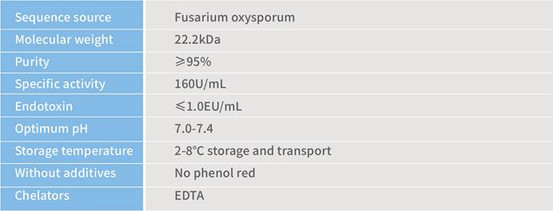
f9a6a5029e6e2c4b330a65d150f2dc72.png


Activity unit: 25℃, pH 7.6, reaction system 3.2ml (1cm optical path). One trypsin unit (USP) is defined as the amount of trypsin that increases absorption value by 0.003 under 253nm through enzymolysis of BAEE per minute.


Applications in Biopharmaceuticals and Cell Research


Hzymes’ recombinant trypsin digestion (TrypLE-like) is versatile across cell culture and bioprocessing workflows:


CHO cell dissociation


Ideal for stable cell lines in protein expression and monoclonal antibody production


HEK293 cells


Reliable for viral vector production, vaccine development, and recombinant protein research


Stem cell dissociation


Gentle and reproducible, supporting embryonic stem cells and iPSC culture


Primary keratinocytes and A549 cells


Preserves morphology and viability for downstream assays


Drug discovery and diagnostics


Facilitates high-quality cell preparation for screening and research


Why Choose Hzymes?


Hzymes is a trusted provider of enzyme solutions for molecular biology, diagnostics, and biopharmaceuticals. With deep expertise in enzyme engineering, GMP manufacturing, and AOF-certified reagents, Hzymes delivers products that enable:


Consistency 


across research and industrial-scale production


Regulatory compliance 


for clinical and manufacturing use


Sustainability 


through elimination of animal-derived components


By choosing Hzymes’ Recombinant Trypsin Digestion (TrypLE-like alternative), researchers and manufacturers gain access to a gentle, easy-to-use, and GMP-grade solution tailored for the future of cell culture.


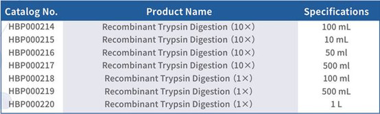
Orderings


7c268a3d43d077d0040ec15fb600e641.png


Conclusion


As cell-based research and biomanufacturing continue to expand, the need for safe, reproducible, and regulatory-compliant reagents grows stronger. Hzymes’ Recombinant Trypsin Digestion (TrypLE-like, GMP Grade) is the ideal choice for scientists and biopharma companies seeking a gentle, AOF, GMP-certified alternative to traditional trypsin.


With proven performance in CHO, HEK293, stem cells, and beyond, it empowers researchers to achieve high-quality results with confidence.

keskiviikko, 19. elokuu 2026

A Validated Endotoxin Detection Workflow for Next-Generation LNP-Delivered mRNA Vaccines

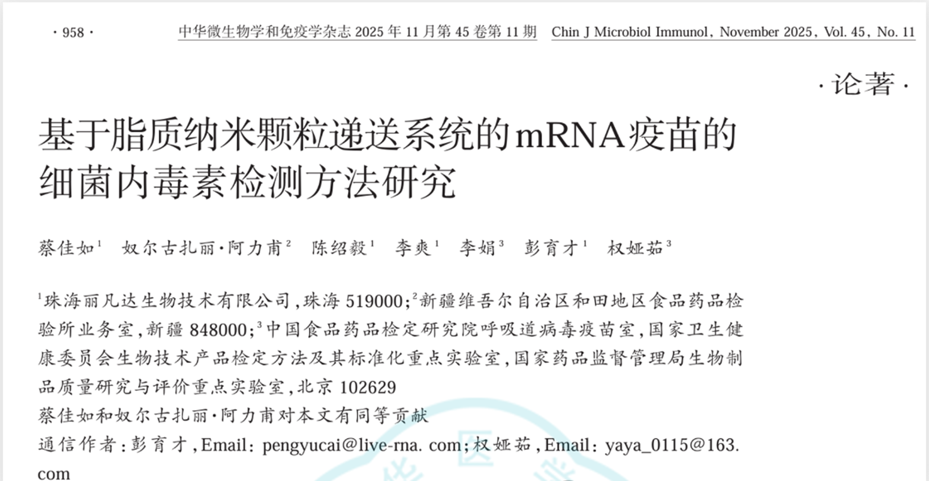
Article Title:


Study on Bacterial Endotoxin Detection Methods for mRNA Vaccines Based on Lipid Nanoparticle Delivery Systems


Published in:


Chinese Journal of Microbiology and Immunology, November 2025


Author Affiliations:


Respiratory Virus Vaccine Division, National Institutes for Food and Drug Control (NIFDC)
Zhuhai Lifanda Biotechnology Co., Ltd.


Published Article


Bacterial Endotoxin Detection Methods Using Recombinant Factor C (rFC) for LNP-mRNA Vaccines


Using the gel-clot limit test described in Part III of the 2020 Chinese Pharmacopoeia, the authors evaluated HSV-2, RSV, and VZV mRNA vaccines. Through optimization of sample dilution factors, diluents, and interference testing, suitable conditions for bacterial endotoxin detection were established.


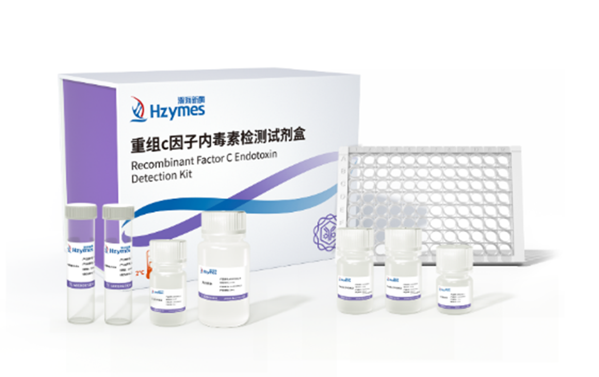
In this study, Hzymes’ Recombinant Factor C (rFC) Endotoxin Assay Kit was used to validate the detection results.


Lipid Nanoparticle (LNP) Structure and Its Impact on mRNA Vaccine Endotoxin Testing


mRNA technology, known for rapid design, high production efficiency, and strong immunogenicity, has opened new avenues in vaccines, oncology, infectious diseases, and autoimmune disorders. The emergency use of COVID-19 mRNA vaccines accelerated its clinical validation and industrialization.


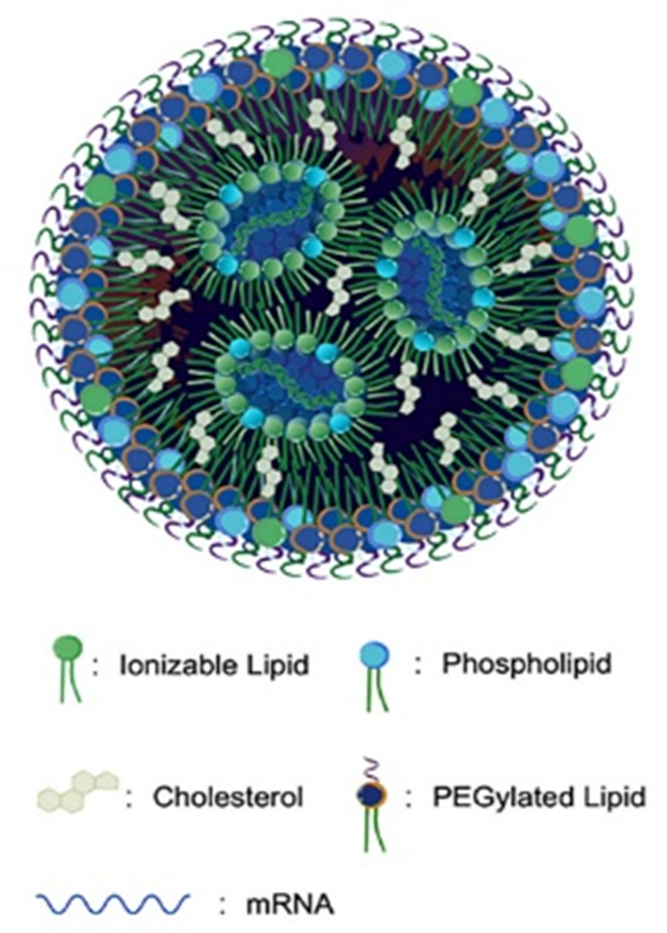
mRNA is delivered into the body via lipid nanoparticles (LNPs), which typically contain cationic lipids, cholesterol, phospholipids, and PEGylated lipids. These components encapsulate nucleic acids through electrostatic interactions.


Structural Diagram of LNP-mRNA


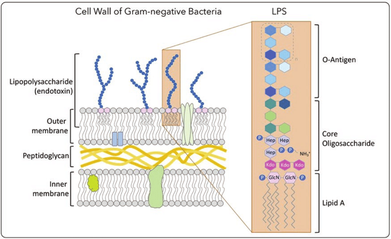
LNP-mRNA vaccines are primarily formulated as injectables, and bacterial endotoxin testing is a critical quality-control requirement. Endotoxins, structural components of Gram-negative bacterial cell walls, consist of O-antigen, core polysaccharide, and lipid A. Even trace amounts (1–5 ng/kg body weight) can trigger severe adverse reactions such as fever, sepsis, DIC, or shock.


Since LNPs are formed by hydrophobic lipid self-assembly, endotoxins may bind to lipid components during detection via electrostatic or hydrophobic interactions. Studies have shown that endotoxins can be adsorbed by liposomes when tested with LAL reagents, causing interference. Diluting lipid concentrations can reduce endotoxin–lipid interactions, and adding divalent cations like magnesium may also suppress interference.


Structural Diagram of Endotoxin


Materials and rFC Testing Methods for Accurate Endotoxin Analysis in LNP-mRNA Formulations


A portion of the study utilized different batches of Hzymes’ rFC assay kits along with unmasking buffer and optimized dilution factors to eliminate LNP-related interference, establish an endotoxin detection method suitable for LNP-mRNA vaccines, and validate the results.


Test Samples


HSV-2, RSV, and VZV mRNA vaccines provided by Zhuhai Lifanda Biotechnology Co., Ltd.


Reagents


Recombinant Factor C assay kits (range: 0.005–5 EU/mL; batch numbers HH20250401R, HH20250101R, HH20240601R) provided by Hzymes Biotech Co., Ltd.


Instrument


SpectraMax i3x fluorescence microplate reader.


Detection Procedures


1.      Using the formula MVD = cL/λ (c = 1.0 mL/mL; L = 30 EU/mL; λ = 0.005 EU/mL), the maximum valid dilution (MVD) was calculated as 6000×. Based on preliminary data, samples were diluted 100× using a Ca/Mg unmasking buffer.


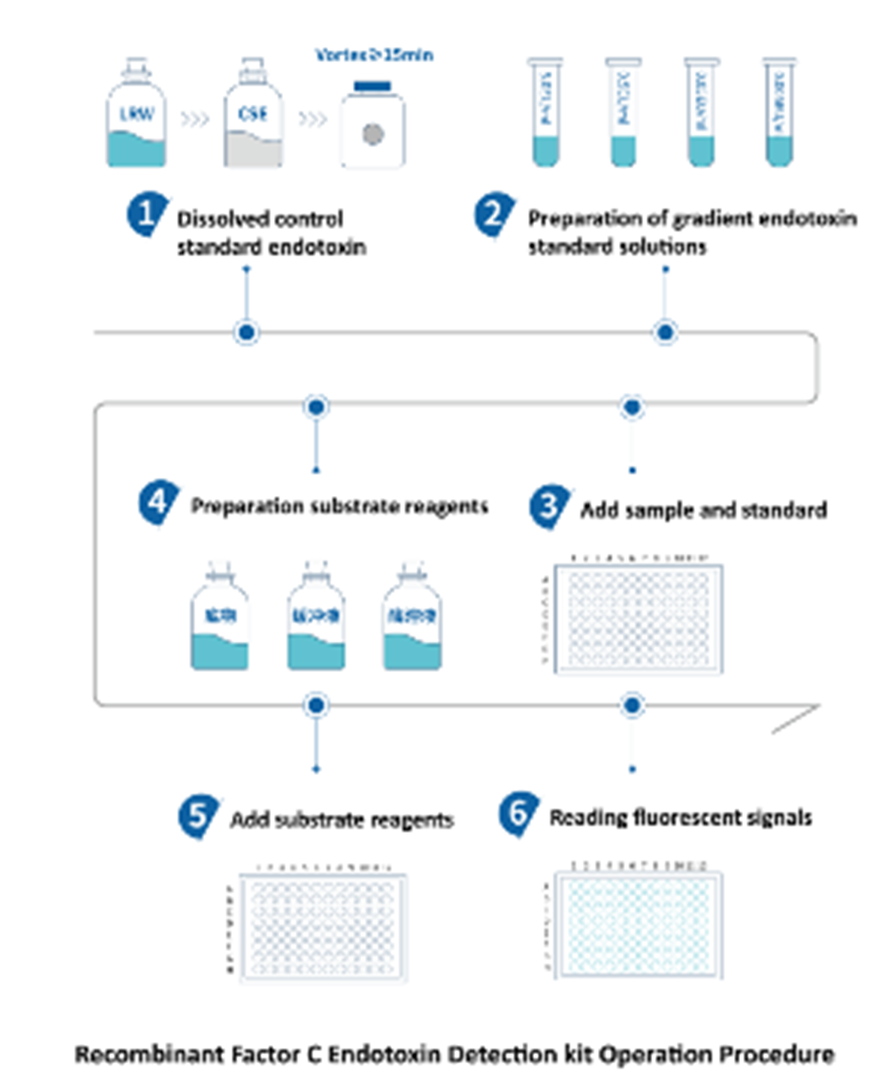
2.      Following the kit instructions, endotoxin standards were prepared at 5, 0.5, 0.05, and 0.005 EU/mL, with endotoxin-free water as the negative control.
Each standard, sample, and control (100 μL) was added to 96-well plates in duplicate, followed by 100 μL of substrate. The instrument was set to Ex 380 nm / Em 440 nm. Fluorescence was recorded at 0 h and after a 1 h incubation at 37°C. ΔRFU was calculated by subtracting initial fluorescence, and net ΔRFU was obtained by subtracting the negative control.


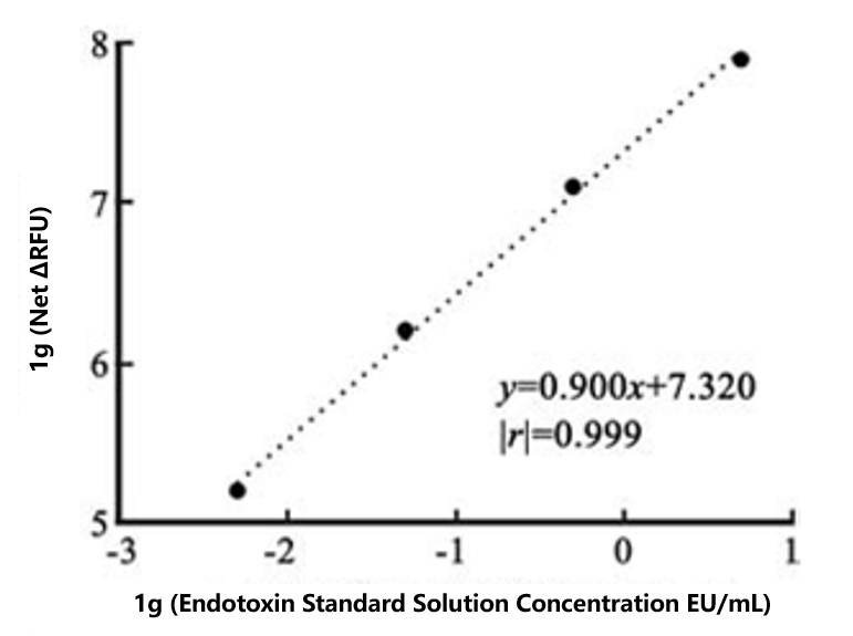
3.      Data Analysis: A linear fit was generated using log concentration (x-axis) and log net ΔRFU (y-axis). Spike recoveries were calculated as:
Recovery (%) = (C_spiked – C_sample) / 0.5 EU/mL × 100%
A recovery of 50–200% indicated no interference.


Validation


1.     Standard Curve Validation:


Three batches of rFC kits produced standard curves with R² = 0.999, 0.990, and 0.999, all ≥0.980, meeting Chinese Pharmacopoeia 2020 requirements. Negative controls were below the lowest standard point (0.005 EU/mL). These curves were therefore valid for endotoxin quantification.


2.     Spike Recovery and Sample Testing:


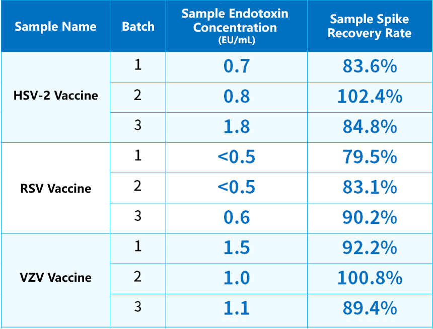
Three batches each of HSV-2, RSV, and VZV vaccines were diluted 100×. One set served as the sample group; the other was spiked to 0.5 EU/mL. Endotoxin concentrations and spike recoveries were calculated using the standard curve equations. Recovery within 50–200% indicated no interference. Qualified samples were then used to calculate final endotoxin content based on the dilution factor.


Endotoxin Testing Results: rFC Assay Performance, Recovery Rates, and LNP Interference Removal


Standard Curve Reliability:


The three kit batches yielded R² values of 0.999, 0.990, and 0.999, all exceeding 0.980. Negative controls remained below the lowest standard point, meeting the validity requirements of General Chapter 1143 (photometric method) in the 2020 Chinese Pharmacopoeia.


Spike Recovery and Detection Results:


Across HSV-2, RSV, and VZV vaccine samples, rFC-based endotoxin measurements showed spike recoveries between 79.5% and 102.4%, within the acceptable 50–200% range. Endotoxin levels were <30 EU/mL and consistent with results obtained by the gel-clot method.


Conclusion and Future Outlook for rFC Endotoxin Assays in LNP-mRNA Vaccine Quality Control


LNP samples may interfere with LAL-based endotoxin assays. Using the unmasking buffer in the rFC kit effectively eliminated this interference. The rFC method produced results consistent with the gel-clot method, supporting its reliability.


This study provides an effective strategy for establishing endotoxin testing methods for LNP-mRNA vaccines, ensuring accuracy by minimizing interference. With the advancement of endotoxin detection technologies, methods such as rFC offer strong complementary value to traditional LAL assays, providing more comprehensive quality-control capabilities for LNP-based mRNA vaccines.


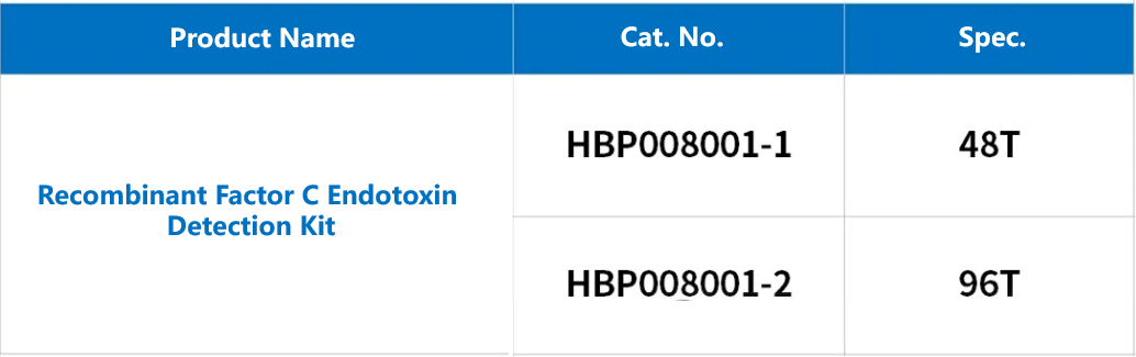
Recombinant Factor C (rFC) Endotoxin Detection Kit – Ordering Information


To obtain the original PDF, please contact: https://www.hzymesbiotech.com/index/user/register?url=/
Please verify: Recombinant Factor C